
Conventional Open Surgery
Learn · Interventional Radiology
Understanding Access to Image-Guided Care.
Explore why interventional radiology matters, how minimally invasive treatment differs from open surgery, and what those differences can mean for a patient’s day.
The Access Problem · 01
Image-Guided Treatment Remains Out of Reach for Many.
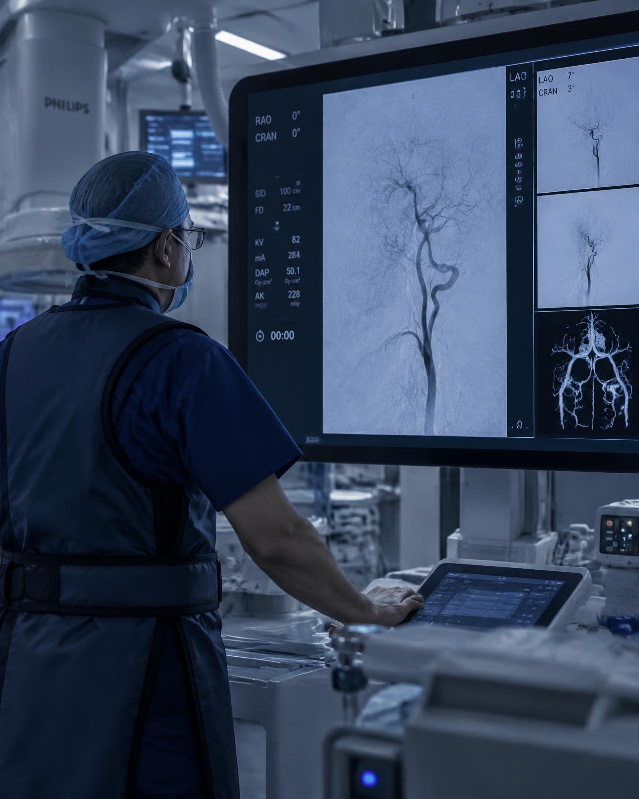
Minimally invasive, image-guided treatment can change the course of a patient’s illness. Access to it still depends heavily on where a patient happens to live, and on whether local services, trained clinicians and referral pathways exist.
Our Vision
A future in which geography does not determine whether a patient can benefit from minimally invasive, image-guided treatment, beginning with strengthening IR capacity in Nigeria and ultimately expanding sustainable, locally led access across West Africa and beyond.
01
Image-Guided Care

02
Access Depends on Where You Are

03
Capacity. People. Pathways.
04
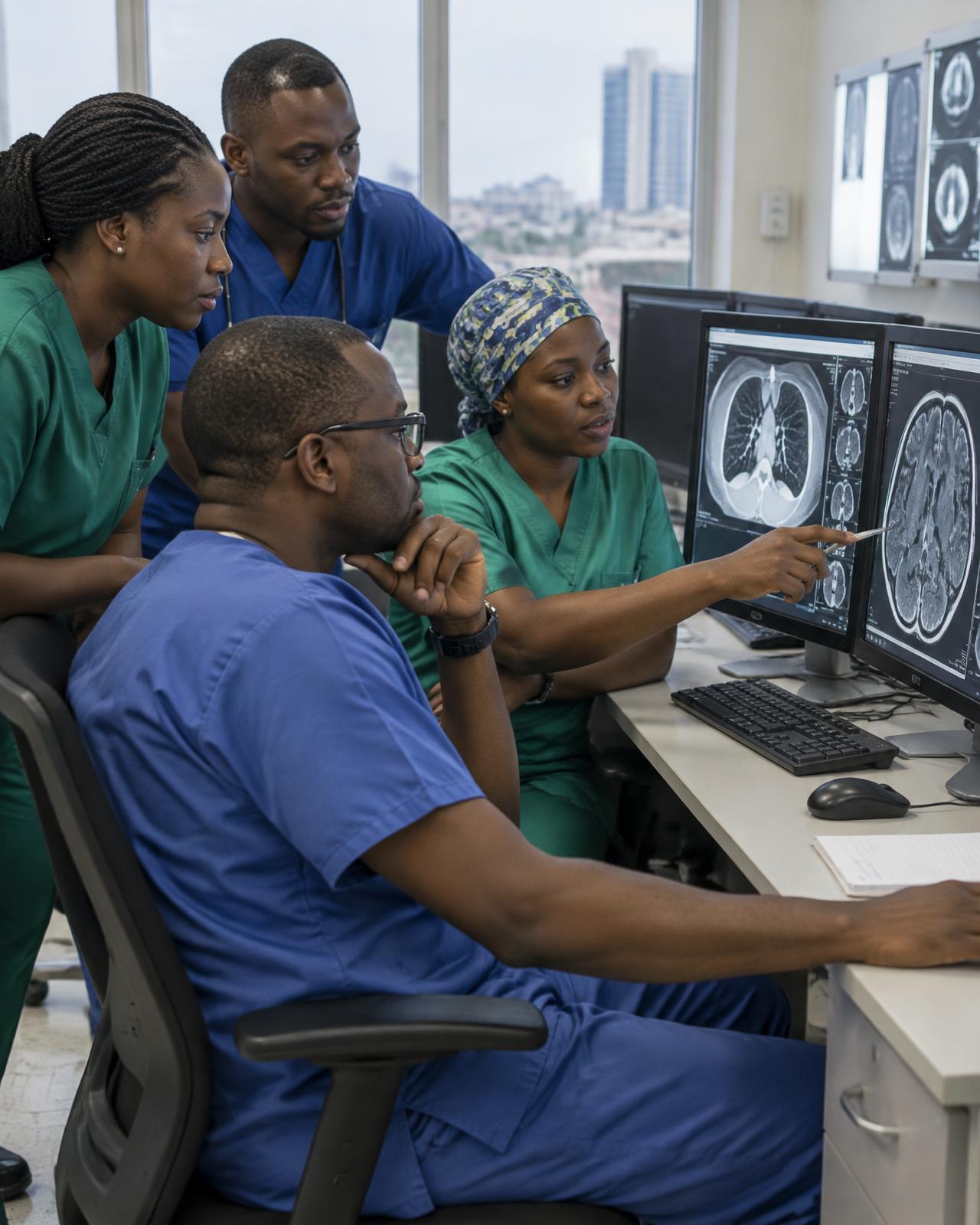
Building Capacity in Nigeria

05
Locally Led. Regionally Connected.
01 / 05
Why It Matters · 02
Two Ways to Treat the Same Problem.
See the stark difference between conventional open surgery and minimally-invasive alternatives.
CHOOSE A CLINICAL SITUATION, THEN COMPARE CONVENTIONAL OPEN SURGERY WITH THE IMAGE-GUIDED ALTERNATIVE ACROSS INCISION, ANAESTHESIA, HOSPITAL STAY AND PHYSIOLOGICAL BURDEN.
Symptomatic uterine fibroids causing heavy bleeding, pain or pressure.
Compare Approach
Minimally Invasive, Image-Guided
Uterine Fibroid Embolisation
Currently Shown
- Incision and Access
Open Surgery
Abdominal incision, typically 10–20 cm, with layered closure and a visible scar.
Image-Guided Treatment
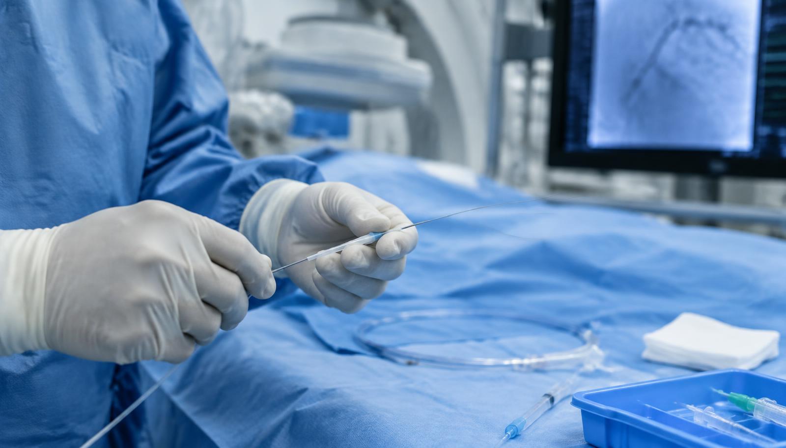
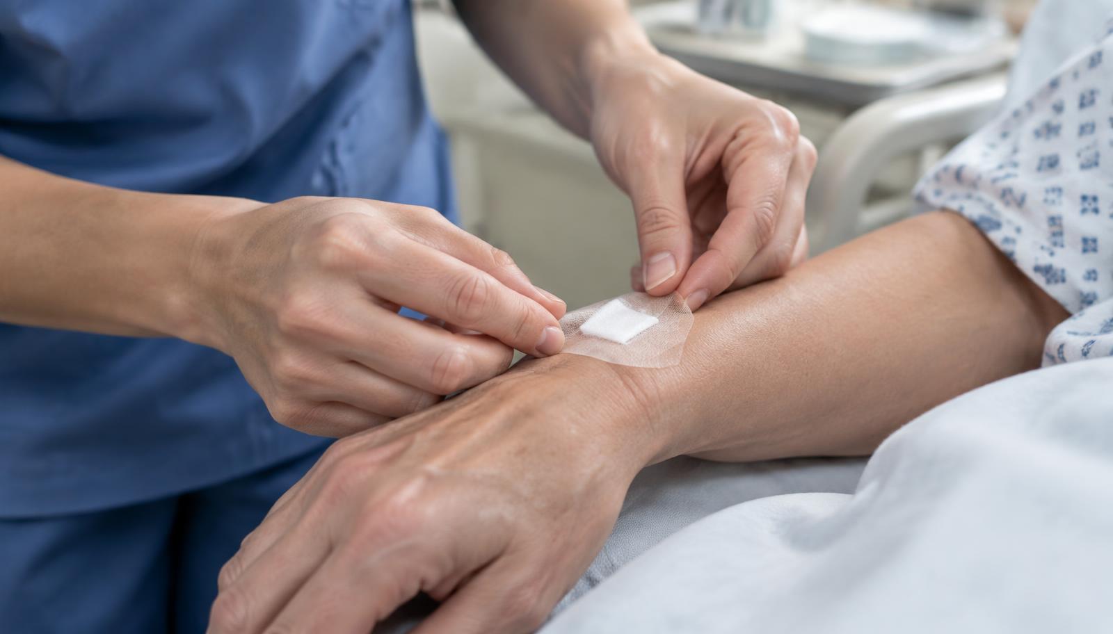
Pinhole puncture at the wrist or groin, a few millimetres wide, closed without stitches.
- Anaesthesia
Open Surgery
General anaesthesia with airway management and an anaesthetic team for the full procedure.
Image-Guided Treatment
Local anaesthesia at the puncture site with conscious sedation and pain control.
- Hospital Stay and Recovery
Open Surgery
Several days as an inpatient, with weeks before a return to usual activity.
Image-Guided Treatment
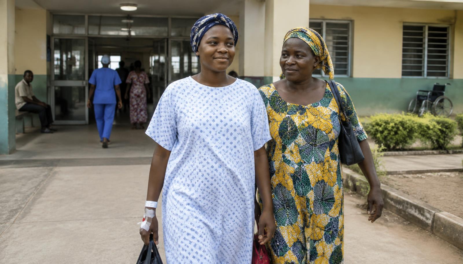
Same-day or overnight observation in most cases, with a shorter return to usual activity.
- Physiological Burden
Open Surgery
Greater blood loss, more wound-related risk and a larger surgical stress response.
Image-Guided Treatment
Targeted treatment through the blood vessels, with the uterus preserved and less blood loss.
Both approaches are legitimate clinical options. Suitability depends on the patient, the imaging findings and the clinical team. This comparison is general clinical information, not medical advice, and not a record of AIR Alliance activity.
A Patient's Day · 03
The Same Problem, Two Very Different Days.
Compare the end-to-end journey for surgical procedures and minimally invasive image-guided treatments through arrival, access, treatment, closure and going home.
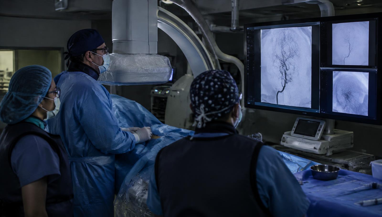
A Day in the Angiography Suite
Follow the same clinical problem through two routes, one open surgery and the other image-guided, from arrival to going home. This is general clinical information, not a record of a specific patient or of AIR Alliance activity. Images are illustrative.
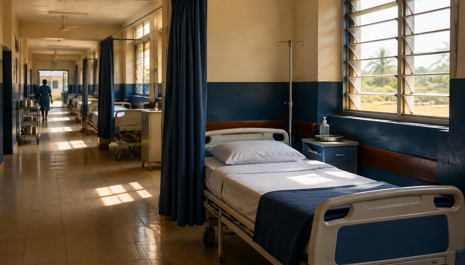 Illustrative photograph of a prepared hospital bed on a quiet ward in the morning
01 / 05
The Physical Footprint, to Scale
Move the control to compare the width of a typical open incision with the width of the puncture used for image-guided access. Both bars are drawn on the same scale.
Open Incision
Approximately 10–20 cm, closed with sutures
Image-Guided Puncture
Approximately 2–3 mm, closed with an adhesive dressing
Sizes are typical ranges given for comparison. The right approach for any patient is decided by their clinical team.