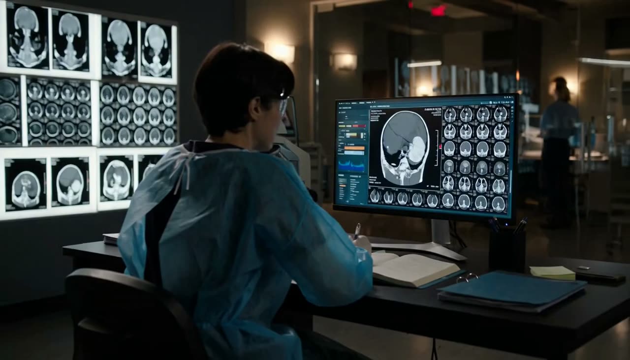
 Illustrative film of an interventional radiology suite
Core · 01
Clinical
Direct clinical care expanding access to minimally invasive, image-guided treatment.
Programmes
CORE organises clinical practice, partnership, evidence and training into a single operating model, and every current programme sits within it.
Illustrative film of an interventional radiology suiteCore · 01
Direct clinical care expanding access to minimally invasive, image-guided treatment.
How the Work Is Organised
Direct clinical practice expanding access to minimally invasive, image-guided interventions, while supporting durable clinical services, workflows and referral pathways.
Building bridges between patients, clinicians, hospitals, professional societies, communities, industry, diaspora physicians and international partners; identifying unmet needs, developing collaborations, raising awareness and connecting resources with locally defined priorities.
Generating and supporting locally relevant research around disease burden, outcomes, barriers to care, appropriate solutions and evidence for policy, investment and clinical practice.
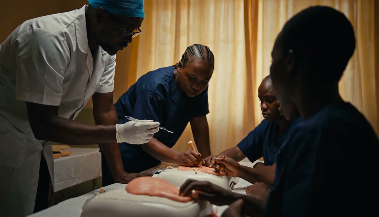
Developing sustainable expertise through specialist training, mentorship, observerships, courses, multidisciplinary education and knowledge exchange.
Current Programmes
Each programme is managed as a first-class record: applications, review, decisions and reporting all run through the same infrastructure.
Support for medical students and radiology residents seeking meaningful exposure to interventional radiology.
Direct interventional radiology care at multiple institutions in Nigeria.
Teaching, mentorship, institutional partnerships and skills development.
Find Your Pathway
Three steps: tell us who you are, where your service or training currently stands, and see the programmes and preparation that apply.
Step 01 · Who You Are
Why Image-Guided Care
CHOOSE A CLINICAL SITUATION Choose a clinical situation, then compare conventional open surgery with the image-guided alternative across incision, anaesthesia, hospital stay and physiological burden.
Symptomatic uterine fibroids causing heavy bleeding, pain or pressure.
Compare Approach
Conventional Open Surgery
Minimally Invasive, Image-Guided
Currently Shown
Open Surgery
Abdominal incision, typically 10–20 cm, with layered closure and a visible scar.
Image-Guided Treatment
Pinhole puncture at the wrist or groin, a few millimetres wide, closed without stitches.
Open Surgery
General anaesthesia with airway management and an anaesthetic team for the full procedure.
Image-Guided Treatment
Local anaesthesia at the puncture site with conscious sedation and pain control.
Open Surgery
Several days as an inpatient, with weeks before a return to usual activity.
Image-Guided Treatment
Same-day or overnight observation in most cases, with a shorter return to usual activity.
Open Surgery
Greater blood loss, more wound-related risk and a larger surgical stress response.
Image-Guided Treatment
Targeted treatment through the blood vessels, with the uterus preserved and less blood loss.
Both approaches are legitimate clinical options. Suitability depends on the patient, the imaging findings and the clinical team. This comparison is general clinical information, not medical advice, and not a record of AIR Alliance activity.
Sponsor a Programme
Support a Training Place or a Clinical Programme.
Giving goes towards the education, clinical support, partnerships and research described on this page.